
Photo credits: Esat Özcan
Thank you, Esat.
- Medial cord
- Radial nerve
- Abductor digiti minimi
- Abductor pollicis brevis
- Abductor pollicis longus
- Adductor pollicis
- Anconeus
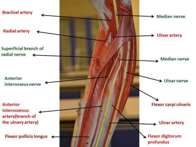
- Anterior interosseous artery (branch of the ulnary artery)
- Anterior interosseus nerve
- Axillary artery
- Bicipital aponeurosis
- Brachial artery
- Brachialis
- Brachioradialis
- Common palmar digital artery
- Common palmar digital nerve
- Coracobrachialis
- Deep branch of radial nerve
- Deep palmar arch
- Deep palmar branch of ulnar artery
- Deltoid
- Dorsal digital artery
- Dorsal digital nerve
- Dorsal interossei
- Dorsal metacarpal artery
- Extensor carpi radialis brevis
- Extensor carpi radialis longus
- Extensor carpi ulnaris
- Extensor digiti minimi
- Extensor digitorum
- Extensor pollicis brevis
- Extensor retinaculum
- Flexor carpi radialis
- Flexor carpi ulnaris
- Flexor digiti minimi brevis
- Flexor digitorum profundus
- Flexor digitorum superficialis
- Flexor pollicis brevis
- Flexor pollicis longus
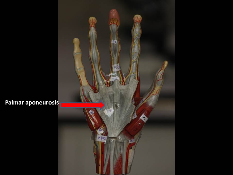
- Flexor retinaculum
- Inferior cutaneous nerve of arm branch of the radial nerve)
- Lateral cutaneous nerve of forearm
- Long head of biceps brachii
- Lumbrical
- Median nerve
- Musculocutaneous nerve
- Opponens digiti minimi
- Opponens pollicis
- Palmar interossei
- Palmar metacarpal artery
- Palmaris longus
- Posterior cutaneous nerve of arm branch of the radial nerve
- Pronator teres
- Proper palmar digital artery
- Proper palmar digital nerve
- Radial artery
- Short head of biceps brachii
- Subscapularis
- Superficial branch of radial nerve
- Superficial palmar arch
- Superficial palmar branch of radial artery
- Supinator
- Tendon of extensor carpi radialis brevis
- Tendon of extensor carpi radialis longus
- Tendon of extensor carpi ulnaris
- Tendon of extensor digit minimi
- Tendon of extensor digitorum
- Tendon of extensor indicis
- Tendon of extensor pollicis brevis
- Tendon of extensor pollicis longus
- Tendon of flexor carpi radialis
- Tendon of flexor digitorum profundus
- Tendon of flexor digitorum superficialis
- Tendon of flexor pollicis longus
- Ulnar artery
- Ulnar nerve
- Axillary nerve
- Circumflex scapular artery
- Infraspinatus
- Lateral head of triceps brachii
- Long head of triceps brachii
- Posterior humeral circumflex artery
- Supraspinatus
- Teres major
- Teres minor
- Triangular space
- Pectoralis major
- Pectoralis minör
- Serratus anterior
- Subclavius
SUPERFICIAL MUSCLES OF THE BACK
- Latissimus dorsi
- Levator scapulae
- Rhomboid major
- Rhomboid minör
- Serratus posterior inferior
- Serratus posterior superior
- Thoracolumbar fascia
- Trapezius
- Adductor brevis
- Adductor canal (Hunter canal)
- Adductor longus
- Adductor magnus
- Adductor minimus
- Femoral artery
- Femoral nerve
- Femoral vein
- Gemellus inferior
- Gemellus superior
- Gluteus maximus
- Gluteus medius
- Gracilis
- Ilıopsoas
- Iliacus
- Iliotibial tract
- Inferior gluteal artery
- Inguinal ligament
- Long head of biceps femoris
- Obturator artery
- Obturator internus
- Obturator nerve
- Pectineus
- Piriformis
- Popliteal artery
- Popliteal vein
- Popliteus
- Profunda femoris artery
- Psoas major
- Quadratus femoris
- Rectus femoris
- Sacral plexus
- Saphenous nerve
- Sartorius
- Sciatic nerve
- Semimembranousus
- Semitendinosus
- Short head of biceps femoris
- Superior gluteal artery
- Tendon of adductor magnus
- Tendon of rectus femoris
- Tensor fascia latae
- Vastus intermedius
- Vastus lateralis
- Vastus medialis
- Abductor digiti minimi
- Abductor hallucis
- Anterior tibial artery
- Calcaneal tendon (Achilles tendon)
- Common dorsal digital nerve
- Common peroneal (fibular) nerve
- Common plantar digital nerve
- Deep peroneal (fibular) nerve
- Deep plantar arch
- Dorsalis pedis artery
- Extensor digitorum brevis
- Extensor hallucis brevis
- Flexor digiti minimi brevis
- Flexor digitorum brevis
- Flexor digitorum longus
- Flexor hallucis brevis
- Flexor hallucis longus
- Inferior extensor retinaculum
- Inferior peroneal retinaculum
- Interossei plantaris
- Lateral head of gastrocnemius
- Lateral plantar artery & nerve
- Lumbrical
- Medial head of gastrocnemius
- Medial plantar artery & nerve
- Peroneus longus
- Peroneus brevis
- Pes anserinus
- Plantar metatarsal artery
- Popliteal vein
- Popliteus
- Posterior tibial artery
- Proper dorsal digital nerve & artery
- Proper plantar digital artery & nerve
- Quadratus plantae
- Soleus
- Superficial peroneal (fibular) nerve
- Superior peroneal retinaculum
- Tendon of extensor digitorum
- Tendon of extensor digitorum longus
- Tendon of extensor hallucis longus
- Tendon of flexor digitorum longus
- Tendon of flexor hallucis longus
- Tendon of tibialis anterior
- Tibial nerve
- Tibialis anterior
- Tibialis posterior
- Transverse head of adductor hallucis


 Yeditepe Anatomy @ Vine