Neurocranium

INDEX

Frontal Bone

  1. Foramen caecum
  2. Frontal crest
  3. Glabella
  4. Nasal part of frontal bone
  5. Nasal process
  6. Orbital part of frontal bone
  7. Squamaous part of  frontal bone
  8. Superciliary arch
  9. Supraorbital margin of  frontal bone
  10. Zygomatic process of  frontal bone

Occipital bone

  1. Basilar part of occipital bone
  2. Cerebellar fossa
  3. Cerebral fossa
  4. Clivus
  5. Condylar canal
  6. External occipital crest
  7. External occipital protuberance
  8. Foramen magnum
  9. Hypoglossal canal
  10. Inferior nuchal line
  11. Internal occipital crest
  12. Internal occipital protuberance
  13. Occipital condyle
  14. Superior nuchal line
  15. Squamous part of occipital bone

Parietal bones

  1. Tuber parietale [Parietal eminence]
    2.  Superior temporal line
  2.   Inferior temporal line

Ethmoid bone

  1. Cribriform (horizontal) plate of ethmoid bone
  2. Crista galli
  3. Middle nasal concha
  4. Orbital plate
  5. Perpendicular plate of  ethmoid bone
  6. Superior nasal concha

Temporal bones

  1. Arcuate eminence
  2. Articular tubercle
  3. Carotid canal
  4. External acoustic opening
  5. Internal acoustic meatus
  6. Jugular foramen
  7. Jugular fossa
  8. Mandibular fossa
  9. Mastoid notch
  10. Mastoid process
  11. Petrous part of temporal bone
  12. Squamous part of temporal bone
  13. Styloid process
  14. Stylomastoid foramen
  15. Tegmen tympani
  16. Trigeminal impression
  17. Zygomatic process of temporal bone

Sphenoid bone

  1. Anterior clinoid process
  2. Carotid groove
  3. Chiasmatic sulcus
  4. Dorsum sellae
  5. Foramen lacerum
  6. Foramen ovale
  7. Foramen rotundum
  8. Foramen spinosum
  9. Greater wing
  10. Hypophysial fossa
  11. Lateral pterygoid plate
  12. Lesser wing
  13. Medial pterygoid plate
  14. Orbital surface of sphenoid bone
  15. Optic canal
  16. Posterior clinoid process
  17. Pterygoid fossa of sphenoid bone
  18. Pterygoid hamulus
  19. Sphenoidal crest
  20. Superior orbital fissure
  21. Tuberculum sellae

SKULL LAB VIDEO IN BOTH LANGUAGES

Antrophological points

Bregma

Glabella

Nasion

Lambdra

Pterion

Major sutures

Lambdoid suture

Coronal suture

Sagittal suture

Instantanatomy

Instagram-Lab in two parts

  Neurocranium-1

ınstanatomy

6 video

Neurocranium-2

ınstanatomy

8 video

Basicranium

figure-1-basicranium2

Skull-Lateral view

figure-4-skull-lateral-view

Occipital bone-Interior view

figure-2-occipital-bone-interior-view

Skull- Inferior view

skull-inferior-view1

Skull-Posterior view

figure-8-skull-posterior-view

Calvaria-Superior view

figure-12-calvaria-superior-view

Skull- Anterior view

figure-8-skull-anterior-view

Perpendicular plate of ethmoid bone – Nasal septum

fig-1-nasal-septum

Superior concha-Middle concha

fig-6-nasal-cavity-lateral-wall

Anterior surface of petrous part of temporal bone

petrous-part-of-temporal-bone-1

Posted in III.Committee | Leave a comment

Vertebral column, ribs, and the sternum

INDEX

Figures in parentheses

  1. Accessory process (13)
  2. Angle of rib (1)
  3. Anterior (pelvic) sacral foramen  (6)
  4. Anterior arch of atlas (4,9)
  5. Anterior tubercle of atlas (9,11)
  6. Anterior tubercle of transverse process (11)
  7. Apex of sacrum  (6)
  8. Auricular surface (7)
  9. Base of the sacrum  (6)
  10. Body of sternum (1)
  11. Cornu sacrale  (7)
  12. Costal cartilage (1,3)
  13. Costal demifacet (for head of rib) (15)
  14. Costal groove (18)
  15. Dens of axis (4,10)
  16. Foramen transversarium (4,5,9,10,11,12)
  17. Head of rib (1,16)
  18. Inferior articular process (5,12,14)
  19. Inferior articular facet /Facet for inf. articular process (14)
  20. Intermediate sacral crest  (7)
  21. Intervertebral disc  (1,4,5)
  22. Jugular notch (3)
  23. Lamina of the vertebral arch (2,5)
  24. Lateral mass of atlas (9)
  25. Lateral sacral crest  (7)
  26. Mammillary process (13)
  27. Manubrium of sternum (1,3)
  28. Median sacral crest  (7)
  29. Pedicle of vertebral arch (11,12)
  30. Posterior arch of atlas (5,9)
  31. Posterior tubercle of atlas (9)
  32. Posterior tubercle of transverse process (11)
  33. Posterior sacral foramen  (7)
  34. Posterior tubercle of atlas (5,9)
  35. Sacral canal (7)
  36. Sacral hiatus  (7)
  37. Sacral promontory  (6)
  38. Spinous process (2,10-12)
  39. Spinous process of axis (5,10)
  40. Spinous process of vertebra prominens (C7) (5,12)
  41. Sternal angle (1,3)
  42. Superior articular facet (7,12)
  43. Superior articular process  (5,6,9,10,11)
  44. Superior articular surfaces of lateral masses (5,9)
  45. Transverse line (s) (6)
  46. Transverse process (1,2, 9-12)
  47. Transverse process of atlas (4,5)
  48. Tubercle of rib (2,16)
  49. Uncinate process (11)
  50. Vertebral body (1. 10-12)
  51. Vertebral body of axis (4,10)
  52. Vertebral foramen (9-12)
  53. Xiphoid process (1)

Instantanatomy

13 instragram videos

Total duration: 2 min 14 seconds

Instagram video #1   15 seconds

Atlas-Superior view: Anterior tubercle of atlas; Posterior tubercle of atlas; Anterior arch of atlas;Posterior arch of atlas; Superior articular surface of lateral masses; Dens of axis

Instagram video #2 13 seconds

Lateral mass of atlas. Cervical vertebra: Vertebral body; Spinous process; Transverse process; Transverse foramen

Instagram video #3  7 seconds

Pedicle of vertebral arch; Lamina of vertebral arch. These two are the two parts of Vertebral arch.

Instagram video #4 7 seconds

Superior articular processes and articular surfaces; Inferior articular processes and articular processes

Instagram video #5  3 seconds

Intervertebral disc; Sacral promontory

Instagram video #6  7 seconds

Head of rib; Neck of rib; Tubercle of rib; Angle of rib

Instagram video #7  12 seconds

Starting with costal groove; Manubrium (of sternum); Body of sternum; Xiphoid process; Sternal angle

Instagram video #8  12 seconds

Lumbar vertebra: Vertebral foramen; Mammillary process; Accessory process. Sacrum: Base of sacrum; Apex of sacrum

Instagram video #9  14 seconds

Sacrum; Sacral canal; Sacral hiatus; Cornu sacrale; Median sacral crest; Intermediate sacral crest; Lateral sacral crest

Instagram video #10 10 seconds

Sacrum: Transverse line; Anterior sacral foramen; Posterior sacral foramen; Auricular surface

Instagram video #11  10 seconds

Cervical vertebra: Anterior tubercle of transverse process; Posterior tubercle of transverse process; Transverse foramen

Instagram video #12  12 seconds

Atlas: Anterior tubercle of atlas; Posterior tubercle of atlas; (longer) Posterior arch of atlas; Anterior arch of atlas; Transverse process of atlas

Instagram video #13  12 seconds

Sternum: Jugular notch; Manubrium; Body of sternum; Xiphoid process Thoracic vertebra: Costal demifacet (for heaf of rib)

Figure 1. Thoracic cage- anterior view

Figure 2. Thoracic cage-posterior view

Figure 3. Sternum

Figure 4. Atlas and axis- anterior view

Figure 5. Atlas and axis-posterior view

Figure 6. Sacrum-anterior view

Figure 7. Sacrum-posterior view

Figure 8.Coccyx

Figure 9. Atlas – Superior view

Atlas superior view

Figure 10. Axis

Axis

Figure 11. Cervical vertebra – A typical one

Cervical vertebra

Figure 12. Vertebra prominens (C7)

Vertebra prominens

Figure 13. Lumbar vertebra

Lumbar vertebra

Figure 14. Thoracic vertebra

Thoracic vertebra

Figure 15. Thoracic vertebra -lateral view

Costal demifacet

Figure 16. Rib (Costa)

Photo credits: Merve Eriş

Rib

Figure 17. Costal groove (Rib -interior view)

Rib-interior view

Figure 18. Anterior and posterior tubercles of transverse process

Ant and post tubercles

Posted in II. Committee | 1 Comment

Bones of the Lower Limb

BONES OF THE LOWER LIMB-INDEX-.PDF VERSION

INDEX

  1. Adductor tubercle (1,3)
  2. Anterior intercondylar area (3)
  3. Apex of head of fibula (4)
  4. Body (shaft) of femur (Corpus femoris) (1)
  5. Calcaneal tuberosity (5)
  6. Calcaneus (4,5,6)
  7. Cuboid (4,6)
  8. Distal  phalanx (5,6)
  9. Facies poplitea (2)
  10. Fibular notch (4)
  11. Fossa intercondylaris (2)
  12. Fovea for the ligament of the head (1)
  13. Gluteal tuberosity (2)
  14. Greater trochanter (Trochanter major) (1,2)
  15. Head of femur (Caput femoris) (1,2)
  16. Head of fibula (4)
  17. Head of talus
  18. Intercondylar eminence (3)
  19. Intermediate cuneiform (4)
  20. Intertrochanteric crest (Crista intertrochenterica) (2)
  21. Intertrochanteric line
  22. Lateral  cuneiform (4,6)
  23. Lateral condyle of femur/Lateral femoral condyle (2,3,10,11)
  24. Lateral condyle of tibia (3,10)
  25. Lateral epicondyle (1,3)
  26. Lateral intercondylar tubercle (3)
  27. Lateral lip  of linea aspera (2)
  28. Lateral malleolus (12)
  29. Lateral supracondylar line (2)
  30. Lateral tubercle (5)
  31. Lesser trochanter (Trochanter minor) (1,2)
  32. Medial  lip of linea aspera (2)
  33. Medial condyle of femur/Medial femoral condyle (2,3,10,11)
  34. Medial condyle of tibia (3,10)
  35. Medial cuneiform (4,5)
  36. Medial epicondyle (1,3)
  37. Medial intercondylar tubercle (3)
  38. Medial malleolus (4,5)
  39. Medial supracondylar line (2)
  40. Medial tubercle (5)
  41. Metatarsal (4,5,6)
  42. Middle phalanx (5,6)
  43. Navicular (4,5,6)
  44. Neck of femur (Collum femoris) (1,2)
  45. Neck of fibula (4)
  46. Neck of talus
  47. Patella(1,3,4)
  48. Patellar surface (1,3)
  49. Pectineal line (2)
  50. Posterior intercondylar area (3)
  51. Proximal phalanx (5,6)
  52. Sustentaculum tali (5)
  53. Talus (4,5,6)
  54. Tibial tuberosity (4)
  55. Trochanteric fossa (Fossa trochenterica) (2)
  56. Trochlea of talus (5)
  57. Posterior articular calceneal surface
  58. Middle articular calcaneal surface
  59. Anterior articular calcaneal surface
  60. Facies articularis navicularis
  61. Posterior articular talar surface
  62. Middle articular talar surface
  63. Anterior articular talar surface
  64. Talar sulcus
  65. Calcaneal sulcus
  66. Lateral malleolar articular surface [Facies malleolaris lateralis]
  67. Medial malleolar articular surface [Facies malleolaris medialis] –in the shape of comma-
  68. Lateral talar process
  69. Fibular trochlea
  70. Navicular tuberosity
  71. Tuberosity of cuboid
  72. Tuberosity of the 5th metatarsal (Styloid process of the 5th metatarsal)
  73. Sesamoid bone of the 1st metatarsal

Instantanatomy

16 instagram videos

Total duration: 3 min 10 seconds

Instagram video #1    12 seconds

Head of femur; Fovea for the ligament of the head; Neck of femur; Greater trochanter; Lesser trochanter

Instagram video #2  14 seconds

Proximal part of femur-posterior view Trochanteric fossa inside the greater trochanter; Intertrochanteric crest. Proximal part of femur-anterior view Intertrochanteric line. Body of femur. Distal part (end) of femur-anterior view Patellar surface

Instagram video #3  11 seconds

Lateral lip of linea aspera continues as Gluteal tuberosity superiorly. Medial lip of linea aspera continues as Pectineal line superiorly. Pectineal line just under lesser trochanter on the posterior view

Instagram video #4     11 seconds

Lateral supracondylar line; Medial supracondylar line; Facies poplitea; Medial condyle of femur; Lateral condyle of femur; cavity between these condyles on the posterior view of distal end of femur Intercondylar fossa

Instagram video #5    14 seconds

Medial epicondyle ; Lateral epicondyle. Epi-means “above”. Adductor tubercle on the medial epicondyle. Proximal end of tibia: Medial condyle of tibia; Lateral condyle of tibia

Instagram video #6        12 seconds

Medial intercondylar tubercle + Lateral intercondylar tubercle = Intercondylar eminence. Anterior intercondylar fossa; Posterior intercondylar fossa

Instagram video #7     12 seconds

Tibial tuberosity; Head of fibula; Apex of head of fibula; Neck of fibula; Medial mallleolus (it means little hammer ; and is at the distal end of tibia); Lateral malleolus (at the distal end of fibula); Fibular notch (on tibia’s distal end. Fibula’s distal end articulates with this notch)

Instagram video #8     11 seconds

Talus: Medial tubercle; Lateral tubercle. Calcaneus under talus. Calcaneal tuberosity; Sustentaculum tali (Little shelf of talus) on the medial side of calcaneus

Instagram video #9        8 seconds

Cuboid bone (anterior to calcaneus on the lateral side); Navicular bone (anterior to talus on the medial side); Medial-intermediate-lateral cuneiform bones

Instagram video #10       7 seconds

Metatarsal; Proximal phalanx; Middle phalanx; Distal phalanx

Instagram video #11        11 seconds

Talus (Ankle bone) Head of talus; Neck of talus; Body of talus. Trochlea of talus

Instagram video #12      13 seconds

Calcaneus – Superior view: Middle articular talar surface on sustentaculum tali; Anterior articular talar surface; Posterior articular talar surface. Between anterior-middle articular talar surfaces and posterior articular talar surface calcaneal sulcus (groove;oluk)

Instagram video #13        14 seconds

Talus-Inferior view: Anterior articular calcaneal surface, Middle articular calcaneal surface; Posterior articular calcaneal surface. Between ant-middle articular calcaneal surfaces & posterior articular calcaneal surfaces calcaneal sulcus (groove; oluk)

Instagram video #14         13 seconds

Talus -Superior view: Facies articularis navicularis (anteriorly located); Trochlea of talus; Lateral talar process

Instagram video #15         14 seconds

Talus- Medial malleolar articular surface (has a shape of a comma); Lateral malleolar articular surface (is triangular and larger). Calcaneus-Lateral view: Fibular trochlea

Instagram video #16      12 seconds

Plantar surface of foot/Inferior view of foot: Navicular tuberosity (on the medial side); Tuberosity of cuboid (on the lateral side); Tuberosity of the 5th metatarsal (Styloid process of the 5th metatarsal); Sesamoid bone of the 1st metatarsal

Figure 1.Femur-anteromedial view

figure-1-femur-anteromedial-view1

Figure 2. Femur-posterior view

figure-2-femur-posterior-view

Figure 3. Femur distal end-Tibia proximal end- Anterior view

figure-3-femur-distal-end-tibia-proximal-end-anterior-view

Figure 4. Right leg & foot-anterior view

figure-4-right-leg-foot-anterior-view

Figure 5. Bones of the foot-medial view

figure-5-bones-of-the-foot-medial-view2

Figure 6. Bones of the foot-lateral view

figure-6-bones-of-the-foot-lateral-view

Figure 7. Lateral malleolus

figure-7-lateral-malleolus

Figure 8. Talus inferior view-Calcaneus superior view

CALCANEUS-TALUS1

Figure 9. Talus inferior view-Calcaneus superior view

TALAR SULCUS

Figure 10. Sinus tarsi: Calcaneal sulcus+Talar sulcus

SINUS TARSI

Figure 11. Inferior surface of talus

Inferior surface of talus

Figure 12.Superior view of talus

Talus-Superior view

Figure 13.Lateral view of talus

Talus-Lateral view

Figure 14.Lateral view of calcaneus

Lateral view of calcaneus

Figure 15. Inferior view of foot

FOOT-PLANTAR SURFACE

Posted in II. Committee | Leave a comment

Nerves & Vasculature of the Lower Limb

INDEX

1 . Watch the 1.5 h video and complete the 4 quizzes! Your score will not be used for academic evaluation!

2. 30-40 min. demonstration at the lab!

3. Then have the V. Committe Lab Exam (1 point) at the Lab on  21.May.2015 Lab on “Nerves & Vasculature of the Lab”.

4. You will draw two questions from two bags. Each question= 0.5 pts.

1st  question from the 1st bag: Show the item (One of the 47 items  in the Index below)

ALL 47 EXCEPT THREE FROM THE MODELS

GREAT SAPHENOUS VEIN, SMALL SAPHENEOUS VEIN & SURAL NERVE FROM CADAVER!

2nd question from the 2nd bag: Which innervates this muscle?

NOT COMPLETING THE 4 QUIZZES

OR YOU’VE COMPLETED THE 4 QUIZZES BUT YOU CHOOSE NOT TO TAKE THE LAB EXAM ON 21.MAY.2015

YOU TAKE THE  V. COMMITTEE LAB EXAM ON 28.MAY.2015

4 QUESTIONS. EACH QUESION 0.25 PTS. 2 ITEMS, 2 WHICH NERVE FOR THIS MUSCLE?

QUESTION BAG 1

bag-of-questions

  1. A. profunda femoris  Figure 1, Vine 6
  2. Anterior tibial artery Figures 14,15, Vine 9, Vine 10
  3. Common dorsal digital nerve Figure 15,Vine 11
  4. Common peroneal (fibular) nerve Figure 6, 7, 8,11, 12, 13,Vine 4
  5. Common plantar digital nerve  Vine 13
  6. Deep peroneal (fibular) nerve Figures 10,15, Vine 10
  7. Deep plantar arch Figure 17,Vine 14
  8. Dorsalis pedis artery Figure 10
  9. Femoral artery Vine 5, Vine 6
  10. Femoral nerve Figures 1,3,5, Vine 2, Vine 5
  11. Femoral vein Figure 1,5, Vine 5
  12. Fibular artery  Figure 14,Vine 9
  13. Genitofemoral nerve Figure 2
  14. Great sapheneous vein  CADAVER FIGURES 10,11,12
  15. Iliohypogastric nerve Figure 2
  16. Ilioinguinal nerve Figure 2
  17. Inferior gluteal artery  Vine 3
  18. Inferior gluteal nerve Figure 4
  19. Inferior lateral genicular artery Figure 8,Vine 7
  20. Inferior medial genicular artery Figure 8,Vine 7
  21. Lateral circumflex femoral artery (Branch of a. profunda femoris)  Vine 6
  22. Lateral femoral cutaneous nerve (Lateral cutaneous nerve of thigh) Figure 2
  23. Lateral plantar artery Figures 16,17,Vine 12
  24. Lateral plantar nerve Figures 16,17, Vine 12
  25. Medial plantar artery Figures 16,17, Vine 12
  26. Medial plantar nerve Figures 16,17,Vine 12
  27. Obturator artery Vine 6
  28. Obturator nerve Vine 2
  29. Plantar metatarsal artery Figure 17,Vine 14
  30. Popliteal artery Figures 7, 8,14 Vine 4, Vine 7, Vine 9, Vine 10
  31. Popliteal vein Figures 7, 8Vine 4
  32. Posterior cutaneous nerve of thigh
  33. Posterior tibial artery Figures 11,13,14,17, Vine 9
  34. Proper dorsal digital artery  Figure 15,Vine 11
  35. Proper dorsal digital nerve Figure 15,Vine 11
  36. Proper plantar digital artery  Figures 16, 17,Vine 13
  37. Proper plantar digital nerve Figures 16,17,Vine 13
  38. Saphenous nerve Figures 1,3,Vine 8
  39. Sciatic nerve  Figures 4, 6, Vine 1, Vine 4
  40. Small sapheneous vein CADAVER FIGURES 13,14
  41. Superficial peroneal (fibular) nerve Figures 9,15, Vine 15
  42. Superior gluteal artery Figure 4, Vine 3
  43. Superior gluteal nerve
  44. Superior lateral genicular artery Figure 8, Vine 7
  45. Superior medial genicular artery Figure 8,Vine 7
  46. Sural nerve (branch of tibial nerve) CADAVER FIGURES 13,15
  47. Tibial nerve Figures 6, 7,8, 11, 12, 13, 14, Vine 4, Vine 10

NERVES & VASCULATURE OF THE LOWER LIMB

LAB DEMONSTRATION VIDEOS

LAB VIDEO 1 – Lumbar plexus- Branches

166 MB

5 min 40 sec

  1. Iliohypogastric nerve
  2. Ilioinguinal nerve
  3. Genitofemoral nerve
  4. Lateral cutaneous nerve of thigh (Lateral cutaneous nerve of thigh)
  5. Femoral nerve
  6. Obturator nerve
  7. + Obturator artery
  8. + Femoral vein
  9. + Femoral artery

QUESTION BAG 2

 theoretical questions bagbag-of-questions

Inferior gluteal nerve == Gluteus maximus

Superior gluteal nerve = Gluteus medius, Gluteus minimus, Tensor fascia latae

Nerve to quadratus femoris: Quadratus femoris, Inferior gemellus

Nerve to obturator internus:  Obturator internus, Superior gemellus

THIGH FOS

Anterior compartment of the thigh

Femoral nerve

Quadriceps femoris (rectus femoris, vastus intermedius, vastus lateralis and vastus medialis)

 Sartorius

Medial compartment of the thigh

Obturator nerve

Adductor longus: 

Adductor brevis: Obturator nerve

Adductor magnus: Obturator nerve (Hamstring part: Tibial nerve)

Gracilis: Obturator nerve

Pectineus: Femoral nerve + Obturator nerve

Posterior compartment of the thigh

Tibial nerve

Semitendinosus& Semimembranosus & Long head of biceps femoris

Common peroneal (fibular) nerve

Short head of biceps femoris  

LEG D. S. T.

Deep fibular nerve

Tibialis anterior

Extensor hallucis longus

Extensor digitorum longus

Superficial fibular nerve

Peroneus (fibularis) longus

Peroneus (fibularis) brevis

Tibial nerve

Gastrocnemius

Soleus

Plantaris

Popliteus

Flexor hallucis longus

Flexor digitorum longus

Tibialis posterior

FOOT

Lateral plantar nerve

All the intrsinc muscles of the foot except the ones below innervated by the medial plantar nerve

Medial plantar nerve

Abductor hallucis, flexor hallucis brevis, flexor digitorum brevis and 1st lumbrical muscle

Deep peroneal (fibular) nerve

Extensor hallucis brevis and extensor digitorum brevis

MODEL IMAGES LABELLED

Figure 1. Posterior abdominal muscles, anterior compartment of thigh

Figure 1

Figure 2. Posterior abdominal muscles and some of the branches of lumbar plexus

Figure 2

Figure 3. Anterior view of thigh and posterior abdominal wall muscles

Figure 3

Figure 4. Gluteal region & Sciatic nerve, Superior & inferior gluteal arteries

Figure 4

Figure 5.  Anterior compartment of the thigh

Figure 5

Figure 6.  Posterior compartment of the thigh

Figure 6

Figure 7.  Posterior compartment of the thigh – deep (long head of biceps femoris and semintendinosus and semimembranposus removed)

Figure 7

Figure 8. Popliteal fossa and branches of popliteal artery

Figure 8

Figure 9. Anterior compartment of the leg

Figure 10

Figure 10. Anterior compartment of the leg- Deep surface

Figure 11

Figure 11. Posterior compartment of the leg- Superficial

Figure 12

Figure 12. Common peroneal nerve and tibial nerve – Superior part of post. compartment of leg : close up

Tibial and common peroneal nerve

Figure 13. Posterior compartment of the leg- Deep

Posterior compartment of leg-Deep

Figure 14. Posterior tibial artery- Fibular artery

posterior-tibial-fibular-artery2

Figure 15. Foot-dorsum

foot-dorsum

Figure 16. Foot- Plantar surface- Superficial 

foot-plantar-surface-superficial

Figure 17. Foot- Plantar surface-Deep [Abductor hallucis muscle moved away]

foot-plantar-surface-3rd-layer-abductor-digiti-hallucis-moved-away

CADAVER IMAGES LABELLED

Cadaver-Figure 1. Gluteal region- deep layer 

Cadaver Figure 1

Cadaver-Figure 2. Posterior abdominal wall

Cadaver Figure 2

Cadaver-Figure 3. Anterior compartment of the thigh

Cadaver Figure 3

Cadaver-Figure 4. Anterior compartment of the thigh- Closer look to the superior part

Cadaver Figure 4

Cadaver-Figure 5. Posterior compartment of the thigh  

Cadaver Figure 5

Cadaver-Figure 6. Medial compartment of the thigh – Inferior part. Adductor canal (Hunter’s canal)

Cadaver Figure 6

Cadaver-Figure 7. Posterior compartment of the thigh

Cadaver Figure 7

Cadaver-Figure 8. Posterior compartment of the leg-Deep surface

Figure 9

Cadaver-Figure 9. Anterior and lateral compartments of the leg- Superficial

anterior-compartment-of-the-leg

Cadaver-Figure 10. Great saphenous vein

great-saphaneous-vein

Cadaver-Figure 11. Great saphenous vein

great-saphaneous-vein-2

Cadaver-Figure 12. Great saphenous vein

great-saphaneous-vein-3

Cadaver-Figure 13. Sural nerve & Small saphenous vein

small-saphaneous-vein

wkgbjlimzviagpqfacmbm85zq4i685

Cadaver-Figure 14.  Small saphenous vein

small-saphaneous-vein-2

Cadaver-Figure 15.  Posterior compartment of the leg- Superficial

posterior-leg-superficial

Vine Lab

15 vine videos = 90 sec= 1.5 min.

VINE 1 

Sciatic nerve

VINE 2

  1. Femoral nerve
  2. Obturator nerve

VINE 3

  1. Superior gluteal artery
  2. Inferior gluteal artery

VINE 4

  1. Sciatic nerve
  2. Tibial nerve
  3. Common peroneal nerve
  4. Popliteal vein
  5. Popliteal artery

VINE 5 (Note: I did click on “report this post” accidentally)

  1. Femoral vein
  2. Femoral artery
  3. Femoral nerve

VINE 6

  1. Obturator artery
  2. Femoral artery
  3. A. profunda femoris
  4. Lateral circumflex femoral artery

VINE 7

  1. Popliteal artery
  2. Superior medial genicular artery
  3. Superior lateral genicular artery
  4. Inferior medial genicular artery
  5. Inferior lateral genicular artery

VINE 8

Saphenous nerve

VINE 9

  1. Popliteal artery
  2. Anterior tibial artery
  3. Posterior tibial artery
  4. Fibular artery (branch of posterior tibial artery)

VINE 10

  1. Deep peroneal nerve
  2. Anterior tibial artery
  3. Posterior tibial artery
  4. Tibial nerve

VINE 11

  1. Common dorsal digital nerve
  2. Proper dorsal digital nerve
  3. Proper dorsal digital artery

VINE 12

  1. Medial plantar nerve
  2. Medial plantar artery
  3. Lateral plantar nerve
  4. Lateral plantar artery

VINE 13

  1. Common plantar digital nerve
  2. Proper plantar digital nerve
  3. Proper plantar digital artery

VINE 14

  1. Deep plantar arch
  2. Plantar metatarsal artery

VINE 15

Superficial peroneal (fibular) nerve

Posted in V. Committee | Leave a comment

Nerves & Vasculature of the Upper Limb

INDEX

  1. A. profunda brachii
  2. Axillary nerve
  3. Brachial artery
  4. Circumflex scapular artery
  5. Deep branch of radial nerve
  6. Lateral cutaneous nerve of forearm
  7. Median nerve
  8. Musculocutaneous nerve
  9. Posterior circumflex humeral artery
  10. Radial nerve
  11. Superficial branch of radial nerve
  12. Ulnar nerve

FIGURES

shoulder-posterior-view

brachial-plexus-2


fig-2-axilla-and-arm-anterior-view

fig-3-upper-limb-anterior-view


forearm-deep

forearm-distal-lateral-view1


fig-5-a-profunda-brachii-and-radial-nerve

fig-6-the-letter-m

lateral-cutaneous-nerve-of-forearm


Posted in IV. Committee | Leave a comment

Joints of the Lower Limb

INDEX

  1. Anterior cruciate ligament (Fig. 4-5)
  2. Anterior tibiofibular ligament (Fig.3)
  3. Fibular (Lateral) collateral ligament (Figs.3-4)
  4. Iliofemoral ligament (Fig. 1)
  5. Ischiofemoral ligament (Fig.2)
  6. Lateral meniscus (Figs.3-5)
  7. Medial meniscus (Fig. 3-5)
  8. Patellar ligament (Fig.3)
  9. Posterior cruciate ligament (Fig. 4-5)
  10. Posterior tibiofibular ligament (Fig. 5)
  11. Pubofemoral ligament (Fig.1)
  12. Quadriceps femoris tendon (Fig.3)
  13. Tibial (Medial) collateral ligament (Figs.3-5)

 LAB VIDEOS ( A TOTAL OF 2 VIDEOS) – in Turkish

NOT A PART OF YOUR LAB EXAM BUT LAB VIDEOS FOR JOINTS OF THE UPPER LIMB (2 videos) – in Turkish

Figure 1. Hip joint-anterior view

Figure 2. Hip joint-posterior view

Figure 3. Knee joint-anterior view

Figure 4 Knee joint-anterior view-tendon removed

Figure 5. Knee joint-posterior view

Posted in III.Committee | Leave a comment

Foot

22. May. 2014 Thursday

14:00- 15:20  GROUP A

15:30- 16:50  GROUP B

4 questions

Each question 0. 3  pts.

A total of 1.2 pts.

First  three  Qs direct questions

Fourth question: Nerve of this muscle

Abductor hallucis, flexor hallucis brevis, flexor digitorum brevis and 1st lumbrical muscle: Medial plantar nerve, all the rest of the muscles of the sole of the foot= Lateral plantar nerve

Extensor hallucis brevis and extensor digitorum brevis= Deep peroneal (fibular) nerve

class notes icon

INDEX

  1. Abductor digiti minimi
  2. Abductor hallucis
  3. Common dorsal digital nerve
  4. Common plantar digital nerve
  5. Deep plantar arch
  6. Extensor digitorum brevis
  7. Extensor hallucis brevis
  8. Flexor digiti minimi brevis
  9. Flexor digitorum brevis
  10. Flexor hallucis brevis
  11. Flexor retinaculum  NOT @ VINE VIDEOS
  12. Inferior extensor retinaculum
  13. Inferior peroneal retinaculum
  14. Lateral plantar artery
  15. Lateral plantar nerve
  16. Lumbrical
  17. Medial plantar artery
  18. Medial plantar nerve
  19. Plantar interossei
  20. Plantar metatarsal artery
  21. Proper dorsal digital artery
  22. Proper dorsal digital nerve
  23. Proper plantar digital artery
  24. Proper plantar digital nerve
  25. Quadratus plantae
  26. Superior extensor retinaculum  NOT @ VINE VIDEOS   (Figure I)
  27. Superficial peroneal (fibular) nerve
  28. Superior peroneal retinaculum
  29. Tendon of flexor digitorum longus
  30. Tendon of extensor hallucis longus
  31. Tendon of flexor hallucis longus
  32. Tendon of tibialis posterior
  33. Transverse head of adductor hallucis

vine icon

Yeditepe Anatomy @ Vine

9 Foot Vine videos

Vine Director: Yasin Delibaş

VINE VIDEO 1

  1. Extensor digitorum brevis
  2. Extensor hallucis brevis
  3. Tendon of extensor hallucis longus

VINE VIDEO 2

  1. Common dorsal digital nerve
  2. Proper dorsal digital nerve
  3. Proper dorsal digital artery

VINE VIDEO 3

  1. Abductor hallucis
  2. Abductor digiti minimi
  3. Flexor digitorum brevis
  4. Quadratus plantae

VINE VIDEO 4

  1. Medial plantar nerve
  2. Medial plantar artery
  3. Lateral plantar nerve
  4. Lateral plantar artery

VINE VIDEO 5  

  1. Tendon of flexor digitorum longus
  2. Lumbrical
  3. Common plantar digital nerve
  4. Proper plantar digital nerve
  5. Proper plantar digital artery

VINE VIDEO 6

  1. Flexor hallucis brevis
  2. Tendon of flexor hallucis longus
  3. Flexor digiti minimi brevis

VINE VIDEO 7

  1. Deep plantar arch
  2. Plantar metatarsal artery
  3. Plantar interossei
  4. Transverse head of adductor hallucis

VINE VIDEO 8

  1. Superficial peroneal (fibular) nerve
  2. Inferior extensor retinaculum
  3. Superior peroneal retinaculum
  4. Inferior peroneal retinaculum

VINE VIDEO 9

Tendon of tibialis posterior

Tendon of flexor digitorum longus

Figure 1. Leg- anterior view

SUPERFICIAL

Figure 2. Leg- anterior view 

DEEP

Figure 3. Leg-posterior view

DEEP

Figure 4. Foot-dorsum

Foot Dorsum

Figure 5 Foot- Plantar surface- Superficial 

Figure 6. Foot- Plantar surface-Deep [Abductor hallucis muscle moved away]

I. Extensor retinacula

EXTENSOR RETINACULA

Posted in V. Committee | 1 Comment

Leg

20. May. 2014 Tuesday

15:00- 16:20  GROUP B

16:30- 17:50  GROUP A

4 questions

Each question 0. 4 pts.

A total of 1.6 pts.

First two Qs direct questions

Third and fourth questions: Nerve of this muscle, function of this muscle

INDEX

  1. Anterior tibial artery 2, 7/ vine icon  46
  2. Calceneal tendon (Achilles tendon) 4 vine icon  2
  3. Deep peroneal (fibular) nerve  2 / I /vine icon,  6
  4. Extensor digitorum longus  1/ I / vine icon 5
  5. Fibular artery 7 vine icon  4
  6. Flexor digitorum longus 6/ VIII/vine icon 3
  7. Flexor hallucis longus  6 / VIII/vine icon 3
  8. Great saphaneous vein  II, III, IV
  9. Lateral head of gastrocnemius muscle 4,5/ VII /vine icon 1
  10. Medial head of gastrocnemius muscle 4 / VII /vine icon 1
  11. Peroneus brevis  3 / I /  vine icon 6
  12. Peroneus longus   3,4 ,6 / I /  vine icon 6
  13. Pes anserinus  6 / IX
  14. Plantaris  5/ VII / vine icon 1
  15. Popliteus  6, 7 vine icon 2
  16. Posterior tibial artery 4,6, 7/ VIII/vine icon 46
  17. Small saphaneous vein  V / VI
  18. Soleus  VIII/vine icon 1
  19. Superficial peroneal (fibular) nerve  1 / I
  20. Sural nerve (branch of tibial nerve) V, VII
  21. Tendon of extensor digitorum longus 1, 2 vine icon 5
  22. Tendon of extensor hallucis longus 1, 2 vine icon  5
  23. Tibial nerve 4,5,6,7 /VII, VIII/vine icon 6
  24. Tibialis anterior   1, 2/ I / vine icon 5
  25. Tibialis posterior 6, 7/VII,VIII/vine icon 2 , 4

FUNCTIONS OF THE MUSCLES

  1.  Tibialis anterior   Dorsiflexes ankle and inverts foot
  2. Extensor hallucis longus Extends great toe and dorsiflexes ankle
  3. Extensor digitorum longus   Extends lateral four digits and dorsiflexes ankle
  4. Peroneus (fibularis) longus Everts foot and weakly plantarflexes ankle
  5. Peroneus (fibularis) brevis Everts foot and weakly plantarflexes ankle
  6. Gastrocnemius Plantarflexes ankle when knee is extended; raises heel during walking; flexes leg at knee joint
  7. Soleus Plantarflexes ankle independent of position of knee; steadies leg on foot
  8. Plantaris Weakly assists gastrocnemius in plantarflexing ankle
  9. Popliteus Weakly flexes knee and unlocks it by rotating femur 5° on fixed tibia; medially rotates tibia of unplanted limb
  10. Flexor hallucis longus Flexes great toe at all joints; weakly plantarflexes ankle; supports medial longitudinal arch of foot
  11. Flexor digitorum longus  Flexes lateral four digits; plantarflexes ankle; supports longitudinal arches of foot
  12. Tibialis posterior Plantarflexes ankle; inverts foot

 INNERVATIONS OF THE MUSCLES   D. S. T.

  1.  Tibialis anterior Deep fibular nerve
  2. Extensor hallucis longus Deep fibular nerve
  3. Extensor digitorum longus Deep fibular nerve
  4. Peroneus (fibularis) longus Superficial fibular nerve
  5. Peroneus (fibularis) brevis Superficial fibular nerve
  6. Gastrocnemius  Tibial nerve
  7. Soleus  Tibial nerve
  8. Plantaris  Tibial nerve
  9. Popliteus  Tibial nerve
  10. Flexor hallucis longus  Tibial nerve
  11. Flexor digitorum longus   Tibial nerve
  12. Tibialis posterior Tibial nerve

class notes icon

vine icon

Yeditepe Anatomy @ Vine

6 Leg Vine videos

Vine director: Aykut UÇAR

Vine video 1 

Plantaris

Lateral head of gastrocnemius

Medial  head of gastrocnemius

Soleus

https://vine.co/v/MgWphYeVUUD

Vine video 2

Calcaneal tendon (Achilles tendon)

Popliteus

Tibialis posterior

https://vine.co/v/MgWT5A75AEa

Vine video 3

Flexor hallucis longus  (Hallucis= Related to big toe)

Flexor digitorum longus

https://vine.co/v/MgWt09DDWh7

Vine video 4

Popliteal artery

Anterior tibial artery

Posterior tibial artery

Fibular artery (branch of posterior tibial artery)

Tibialis posterior muscle

https://vine.co/v/MgWUurFQH9Y

Vine video 5

Tibialis anterior

Extensor digitorum longus

Tendon(s) of extensor digitorum longus

Tendon of extensor hallucis longus

https://vine.co/v/MgWx19mB391

Vine video 6

Peroneus longus

Peroneus brevis

Deep peroneal nerve

Anterior tibial artery

Posterior tibial artery

Tibial nerve

https://vine.co/v/MgWQet6Ap9m

Figure 1. Anterior compartment of the leg- Superficial

leg-anterior-superficial

Figure 2. Anterior compartment of the leg – Deep

leg-anterior-deep1

Figure 3.  Lateral compartment of the legleg-lateral-view

Figure 4.  Posterior compartment of the leg- Superficialleg-posterior-superficial1

Figure 5. Plantaris muscle

Plantaris

Figure 6. Posterior compartment of the leg- Deep

leg-posterior-deep3

 Figure 7. Posterior tibial artery & Fibular artery

Posterior tibial fibular artery

Figure I. Anterior and lateral compartments of the thigh- Superficial

Anterior compartment of the thigh

Figures II.,III. IV.  Great saphaneous vein

Great saphaneous vein

Great saphaneous vein-2

Great saphaneous vein-3

Figures V.,VI. Small saphaneous vein

Small saphaneous vein

Small saphaneous vein-2

Figure VII.  Posterior compartment of the thigh- SuperficialPosterior thigh superficial

71

Sural-Nerve-580x676

wKgBJlIMZViAGPqfACMBm85Zq4I685

 Figure VIII.  Posterior compartment of the thigh- Deep

Posterior thigh deep

Figure IX. Pes anserinus

Pes anserinus

Posted in V. Committee | Leave a comment

Thigh & Popliteal fossa

15. May. 2014 Thursday

15:00- 16:20  GROUP A

16:30- 17:50  GROUP B

4 questions

Each question 0. 4 pts.

A total of 1.6 pts.

First two Qs direct questions

Third and fourth questions: Nerve of this muscle, function of this muscle

LAB GUIDE

LAB REHBERİ

class notes icon

INDEX

  1. A. profunda femoris (2) vine icon 9
  2. Adductor brevis (under adductor longus)  vine icon  2
  3. Adductor canal (Hunter’s canal)  (2) / IV vine icon  4
  4. Adductor longus (1,2) / I, II / vine icon 2
  5. Adductor magnus (3,4,5) vine icon  6
  6. Adductor minimus (4) vine icon 6
  7. Common peroneal (fibular) nerve (3,4,5) / III, V vine icon 7
  8. Femoral artery (1,2) / I, II, IV  /vine icon  3 , 9
  9. Femoral nerve (1,2) /  II  / vine icon 3
  10. Femoral vein (1,2) vine icon 3
  11. Gracilis (2,3,5) vine icon 3 , 5
  12. Iliopsoas (1) / I / vine icon 2
  13. Inferior lateral genicular artery (6) vine icon 8
  14. Inferior medial genicular artery (6) vine icon 8
  15. Lateral circumflex femoral artery (Branch of a. profunda femoris) vine icon 9
  16. Long head of biceps femoris (3,6) / III, V vine icon 6
  17. Obturator artery vine icon 9
  18. Patellar ligament (2) vine icon 4
  19. Pectineus (2) / I, II / vine icon 2
  20. Popliteal artery (5,6) vine icon 7 , 8
  21. Popliteal vein (5,6) vine icon 7
  22. Rectus femoris (1) / I vine icon 1
  23. Saphenous nerve (2) / IV vine icon 4
  24. Sartorius (1,3) / I,II, IV vine icon 3  , 5
  25. Sciatic nerve (4,5) / III, V vine icon 7
  26. Semimembranosus (3) / III, V vine icon 5
  27. Semitendinosus (3) / III, V vine icon 5
  28. Short head of biceps femoris (5)/ V vine icon 6
  29. Superior lateral genicular artery (6) vine icon 8
  30. Superior medial genicular artery (6) vine icon 8
  31. Tendon of adductor magnus (2) vine icon 4
  32. Tendon of rectus femoris (2) vine icon 4
  33. Tibial nerve (3,4,5,6) / III, V vine icon 7
  34. Vastus intermedius (2) vine icon 1
  35. Vastus lateralis (2) / I, II vine icon  1
  36. Vastus medialis (2) / I, IV vine icon 1

FUNCTIONS OF THE MUSCLES

Anterior compartment of the thigh

Ilipsoas: Chief flexor of the thigh

Quadriceps femoris: Extension of the knee, extension of the leg at knee joint Rectus femoris also steadies hip joint and helps iliopsoas flex thigh

Sartorius: Flexes, abducts, and laterally rotates thigh at hip joint; flexes leg at knee joint (medially rotating leg when knee is flexed)

Medial compartment of the thigh

Adductor longus: Adducts and medially rotates thigh at hip joint

Adductor brevis: Adducts thigh at hip joint

Adductor magnus: Adducts and medially rotates thigh at hip joint

Gracilis: Adducts thigh; flexes leg; helps rotate leg medially

Pectineus: Adducts and flexes thigh; assists with medial rotation of thigh

Posterior compartment of the thigh

Semitendinosus& Semimembranosus:

Extend thigh; flex leg and medially rotate thigh and leg; when thigh and leg are flexed, these muscles can extend trunk

Biceps femoris:  Flexes leg and rotates it laterally when knee is flexed; extends thigh (e.g., accelerating mass during first step of gait).

 INNERVATIONS OF THE MUSCLES

Anterior compartment of the thigh

Quadriceps femoris: Femoral nerve

Sartorius: Femoral nerve

Medial compartment of the thigh

Adductor longus: Obturator nerve

Adductor brevis: Obturator nerve

Adductor magnus: Obturator nerve (Hamstring part: Tibial nerve)

Gracilis: Obturator nerve

Pectineus: Femoral nerve

Posterior compartment of the thigh

Semitendinosus& Semimembranosus & Long head of biceps femoris (Hamstring muscles) : Tibial nerve

Short head of biceps femoris:  Common peroneal (fibular) nerve

vine icon

Yeditepe Anatomy @ Vine

9 Thigh & Popliteal Fossa Vine videos

Vine director: Aykut UÇAR

Vine video 1

Quadriceps femoris muscle is formed by:

Rectus femoris

Vastus lateralis

Vastus medialis

Vastus intermedius

https://vine.co/v/MrXVWuF75aQ

Vine  video 2

Ilipsoas

Pectineus

Adductor longus

Adductor brevis

https://vine.co/v/MrXY1Pb3zin

Vine video 3  (Note: I did click on “report this post” accidentally)

Sartorius

Gracilis

Adductor magnus

Femoral vein

Femoral artery

Femoral nerve

https://vine.co/v/MrHZFA2uJO2

Vine video 4

Tendon of rectus femoris

Tendon of adductor magnus

Adductor canal

Saphenous nerve

https://vine.co/v/MrHA1a6DvOV

Vine video 5

Sartorius

Gracilis

Semitendinosus

Semimembranosus

https://vine.co/v/MrHqTjhPZT5

Vine video 6

Long head of biceps femoris

Short head of biceps femoris

Adductor minimus

Adductor magnus

https://vine.co/v/MrHhvqMEBYU

Vine video 7

Sciatic nerve

Tibial nerve

Common peroneal nerve

Popliteal vein

Popliteal artery

https://vine.co/v/MrHMJhtX7Xe

Vine video 8

Popliteal artery

Superior medial genicular artery

Superior lateral genicular artery

Inferior medial genicular artery

Inferior lateral genicular artery

https://vine.co/v/MrHem5MxwvA

Vine video 9

Obturator artery

Femoral artery

A. profunda femoris

Lateral circumflex femoral artery

https://vine.co/v/MrHJeIQPxYF

 Figure 1. Anterior compartment of the thigh

Figure 2. Anterior and medial compartments of the thigh (Rectus femoris removed)

anterior-thigh

medial-thigh-musclesmedial-thigh-2

Figure 3. Posterior compartment of the thigh

IMPORTANT NOTE: The model is confusing for semitendinosus and semimembranosus. If I ask, I will ask these muscles from the cadaver. Please see the Figure V.

medial-thigh-musclesmedial-thigh-muscles

Figure 4. Adductor minimus (Superior part of adductor magnus)

Adductor minimus

adductor-magnus185adductor-magnus185 adductor_magnus1332256393104-14152D06A314CE32EAB

Figure 5. Posterior compartment of the thigh – deep (long head of biceps femoris and semintendinosus and semimembranposus removed)

Deep posterior thigh

Figure 6. Popliteal fossa and branches of popliteal artery

Popliteal fossa

 I. Anterior compartment of the thigh

Anterior thigh

II.  Anterior compartment of the thigh- Closer look to the superior part

Anterior thigh-close

III.  Posterior compartment of the thigh  Posterior thigh

IV.  Medial compartment of the thigh – Inferior part. Adductor canal (Hunter’s canal)

Adductor canal

V.  Posterior compartment of the thigh – Short head of biceps femoris Short head of biceps femoris


Posted in V. Committee | 1 Comment

Abdominal muscles-Lumbosacral Plexus- Gluteal region

30-APRIL-2014   WEDNESDAY

14:00- 15:20  GROUP B

15:30- 16:50  GROUP A

Questions

0.2 pts. each Question

TOTAL 0.6 POINTS

INDEX

  1. Aponeurosis of external oblique (6) V
  2. External oblique II, IV (6) V
  3. Femoral nerve (1,4) III (Vine video:6)
  4. Genitofemoral nerve (3) III
  5. Gluteus maximus  I  (Vine video: 2)
  6. Gluteus medius (5)  (Vine video:2)  (Vine video:4)
  7. Iliacus (1,2,3,4) III (Vine video:5)
  8. Iliohypogastric nerve (3)
  9. Ilioinguinal nerve (3)
  10. Iliopsoas (1,4) (Vine video:5)
  11. Iliotibial tract (7) VI
  12. Inferior gemellus  (5) (Vine video:3)
  13. Inferior gluteal artery I (5) (Vine video:4)
  14. Inguinal ligament (1,4,7)
  15. Internal obliue II, IV (6)
  16. Intersection tendon (6) V (Vine video:1)
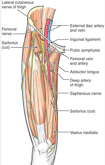
  17. Lateral femoral cutaneous nerve (Lateral cutaneous nerve of thigh) (3) III, VI
  18. Linea alba  (6) (Vine video:1)
  19. Obturator internus  I (5) (Vine video:3)
  20. Obturator nerve (Vine video:6)
  21. Piriformis  I (5) (Vine video:2) (Vine video:3) (Vine video:4)
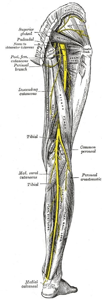
  22. Posterior cutaneous nerve of thigh VII
  23. Psoas major (1,2,3,4) III (Vine video:5) (Vine video:6)
  24. Pyramidalis (6)
  25. Quadratus femoris (5) (Vine video:3)
  26. Quadratus lumborum (3) III
  27. Rectus abdominis (6) V (Vine video:1)
  28. Rectus sheath (6) II (Vine video:1)
  29. Sacral plexus (1)
  30. Saphenous nerve (4)
  31. Sciatic nerve  I (Vine video:2)
  32. Superior gemellus I (5) (Vine video:3)
  33. Superior gluteal artery (5)  (Vine video:4)
  34. Tendinous intersection (6)
  35. Tensor fasciae latae (1,7) VI
  36. Transversus abdominis IV

Cadaver figures demonstrated first by Roman numerals.

Muscles & Fxns:

External oblique: 1. Compresses and supports abdominal viscera 2. Flexes and rotates trunk

Gluteus maximus: 1. Powerful extensor of (especially) flexed femur at hip joint 2. Lateral stabilizer of hip joint and knee joint 3.Laterally rotates and abducts thigh 4.steadies thigh 5.Assists in rising from sitting position

Gluteus medius: 1. Abducts and medially rotates thigh 2. Keeps pelvis level when ipsilateral limb is weight-bearing 

Iliopsoas: Chief flexor of the thigh

Inferior gemellus: 1. Lateral rotation of the thigh, 2.Abduction of the flexed thigh @ hip joint 3. Steady femoral head in acetabulum

Internal obliue: 1. Compresses and supports abdominal viscera 2. Flexes and rotates trunk

Obturator internus: 1. Lateral rotation of the thigh, 2.Abduction of the flexed thigh @ hip joint 3. Steady femoral head in acetabulum

Piriformis 1. Lateral rotation of the thigh, 2.Abduction of the flexed thigh @ hip joint 3. Steady femoral head in acetabulum

Pyramidalis Tenses linea alba

Quadratus femoris: 1. Lateral rotation of the  thigh 2. Steady femoral head in acetabulum

Quadratus lumborum 1. Depresses and stabilizes rib XII during inspiration 2.  Some lateral flexion of trunk

Superior gemellus: 1. Lateral rotation of the thigh, 2.Abduction of the flexed thigh @ hip joint 3. Steady femoral head in  acetabulum

Rectus abdominis: Compress abdominal contents; flexes vertebral column (spine flexion); tense abdominal wall

Tensor fasciae latae: Stabilizes the knee in extension

Transversus abdominis: Compresses and supports abdominal viscera

Muscles & Innervations:

External oblique: T7-T11 spinal nerves and subcostal nerve

Gluteus maximus: Inferior gluteal nerve

Gluteus medius: Superior gluteal nerve

Iliopsoas: Psoas major by the anterior rami of lumbar nerves L1, L2, L3 and Iliacus by femoral nerve

Inferior gemellus : Nerve to quadratus femoris

Internal obliue:Anterior rami of T6-T12 spinal nerves) and first lumbar nerves

Obturator internus: Nerve to obturator internus

Piriformis: Nerve to piriformis

Pyramidalis: Anterior ramus of T12

Quadratus femoris: Nerve to quadratus femoris

Quadratus lumborum: Anterior branches of T12 and L1-L4 nerves

Superior gemellus: Nerve to obturator internus

Rectus abdominis: Anterior rami of T6-T12 spinal nerves

Tensor fasciae latae: Superior gluteal nerve

Transversus abdominis:Anterior rami of T6-T12 spinal nerves) and first lumbar nerves

vine icon

Yeditepe Anatomy @ Vine

Vine for anatomy

        Vine Director:  Aykut UÇAR

VINE VIDEO 1: Abdominal muscles -1

  1. Rectus sheath
  2. Rectus abdominis
  3. Intersection tendon
  4. Linea alba

Vine: https://vine.co/v/MnL96IWWuFz

VINE VIDEO 2: Gluteal region-1

1. Gluteus maximus

2. Gluteus medius

3. Piriformis

4. Sciatic nerve

Vine: https://vine.co/v/MnLder3ghAW

VINE VIDEO 3: Gluteal region-2

1. Piriformis

2. Superior gemellus

3. Obturator internus

4. Inferior gemellus

5. Quadratus femoris

Vine: https://vine.co/v/MnLp0KddvBP

VINE VIDEO 4: Gluteal region-3

1. Gluteus medius

2. Piriformis

3. Superior gluteal artery

4. Inferior gluteal artery

Vine: https://vine.co/v/MnLDvmvE5bw

VINE VIDEO 5:  Gluteal region-5

  1. Psoas major
  2. Iliacus
  3. Iliopsoas

Vine: https://vine.co/v/MnLIjE6QW6O

VINE VIDEO 6: Gluteal region-6

  1. Psoas major
  2. Femoral nerve
  3. Obturator nerve

Vine: https://vine.co/v/MnLTPtO73LK

  MODEL FIGURES

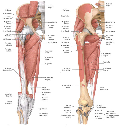
Figure 1. Posterior abdominal wall muscles, femoral nerve and tensor fascia latae

anterior-medial-compartments-of-the-thigh-deep1

Figure 2. Iliacus and psoas major  iliacus-and-psoas-major

Figure 3. Posterior abdominal muscles and some of the branches of lumbar plexus

Posterior abdominal wall muscles & Lumbar plexus

Figure 4. Anterior view of thigh and posterior abdominal wall muscles

Anterior view of thigh and post.abdominal wall muscles

Figıre 5. Gluteal region

Gluteal region

Figure 6. Anterolateral abdominal wall

Anterolateral abdominal wall


Figure 7. Tensor fascia latae – Iliotibial tract

Tensor fascia latae - Iliotibial tract

 CADAVER FIGURES 

Figure I. Gluteal region- deep layer

Cadaver1- Gluteal region

Figure II. Anterolateral abdominal wall

Cadaver-2 Anterolateral abdominal wall

Figure III. Posterior abdominal wall

Cadaver-2 Posterior abdominal wall

Figure IV. The three flat mucles of the anterolateral abdominal wall

Cadaver-4Figure V. Rectus abdominis

Rectus abdominis

Figure VI. Tensor fascia latae


Cadaver VI


Figure VII. Posterior cutaneous nerve of thigh

Gray83220140425_143430

Posted in V. Committee | Leave a comment